SUMMARY(anat15)
![]()
Cardiovascular system 心臟循環器系
|
1: Pectoral fin |
|
1. Ventricle |
1. Heart
2. Vascular system
3. Lymphatic system
Heart 心臟
--- Cranioventral end of the pectoral girdle
--- Located in the pericardial cavity
--- Location
--- Just posterior to gill and to some extent ventral to gill
--- Anterior to the abdominal cavity
|
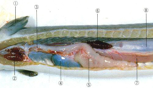
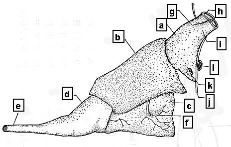
A left-sided view |
|
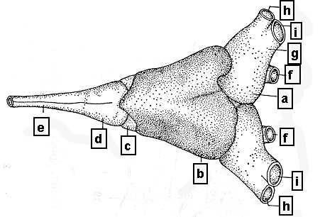
A dorsal view |
1. Compostion of heart
|
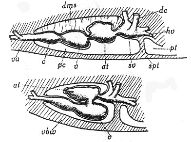
Embryonic stage dc: Cuvier duct, hv: hepatic vein, dms: dorsal mesentry, pt: peritonium, |
|
Adult stage |
--- S-shaped structure of the heart
--- a. Venous sinus (Sinus venosus)
b. (Cardiac) atrium (Atrium cordis)
c. (Cardiac) ventricle (Ventriculum dordis)
d. Cardiac bulb (Bulbus cordis)
--- Originated from yolk-sac veins (Vitelline veins, Venae vitellinae)
--- Cardia ventricle\ cardiac bulb
--- A thickening of the muscular wall of the heart tube
--- Species variation in carciac anatomy
--- Layers of the heart
--- a. Epicardium with subepicardium
b. Myocardium
c. Endocardium with subendocardium
1) Venous sinus
--- Located in dorso-caudal region of pericardial cavity
(1) Common cardinal veins
--- Bilaterally located
--- Running down along both sides of esophagus
(2) Hepatic veins
--- Generally posterior wall of the sinus through the penetration of diaphragm
(3) Jugular and subclavian veins
2) Sino-atrial opening and sinoatrial valve (SA valve)
--- Folding both walls of the sinus and atrium
3) (Cardiac) atrium
--- Dorsal to ventricle and even the cardiac bulb
--- Cardiac auricle 心耳
--- Frequent ventral protrusions of atrium
Coronary sulcus 冠狀溝
--- Furrow between the atrium and ventricle
4) Atrio-ventricular opening
|
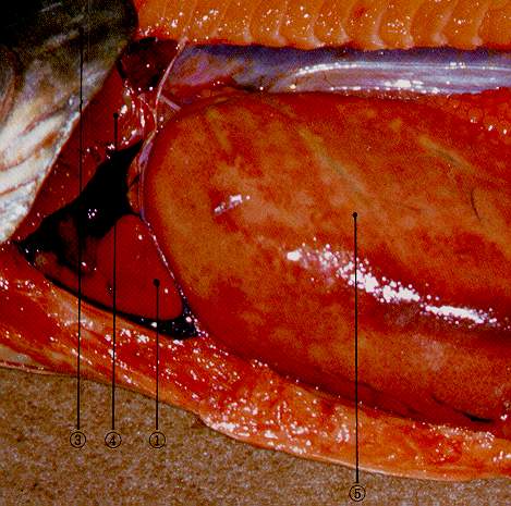
Atrium and ventricle of carp |
--- Always located in dorsal wall of the ventricle
--- Usually 2 equal valves
5) (Cardiac) ventricles
--- Round type/ Triangular type
--- Spongy layer/ Compact layer
6) Arterial opening
--- Between the cardiac ventricle and cardiac bulb
7) Cardiac bulb 心臟球
--- Species variation
---
Arterial cone
--- Original form of cardiac bulb found in shark and ray
--- Separated from the other heart divisions
---
Many longitudinal rows of valves on internal surface (Fig. 189)
Aortic bulb in many teleosts
--- Regression from cardiac bulb
--- Functions
--- Maintenence of smooth blood flow leading to prevention of overpressure load to gill
8) Arterial trunk (Ventral aorta) 動脈幹
--- The first portion of the arterial vascular system except for teleostei
--- a. Arterial cone in primitive teleosts 動脈圓錐
b. Arterial or aortic bulb only in teleostei
9) Ventral aorta (AOv)
|
|
|
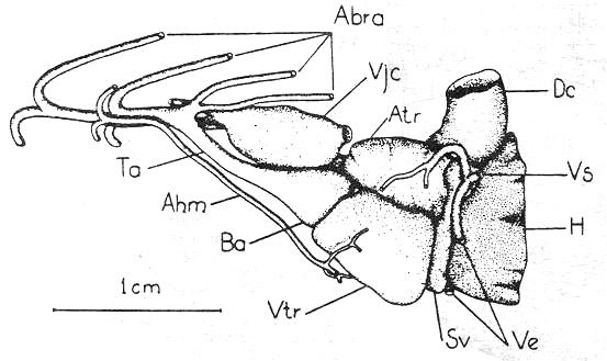
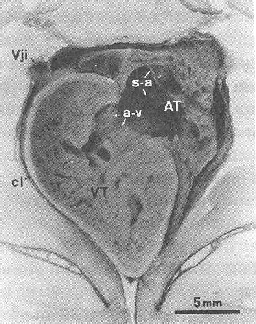
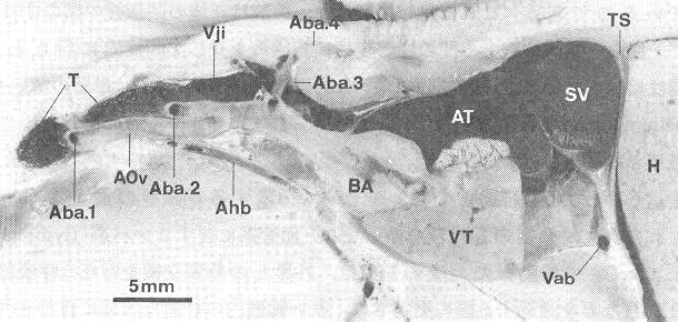
T: Thyroid gland, Aba.1 - 4: The first to 4th afferent branchialarteries, Vji: Inferior jugular vein, Ahb: Hypobranchial artery, AOv: Ventral aorta, BA: Aortic bulb, AT: Atrium, VT: Ventricle, SV: Venous sinus, Vab: Abdominal vein, TS: Transverse septum, H: Liver |
10) Pericardial cavity
![]()
Nutrient vessels of the heart
1) Arterial supplies
--- Dorsal aorta\ Last efferent branchial arteries
2) Venous system
--- Coronary sinus\ Capillary-luminar anstomosis in higher vertebrates
(1) Cardiac bulb
--- Anterior cardinal vein
(2) Ventricular wall
--- Pouring into atrial cavity near atrio-ventricular opening (Ostium atrioventriculare)
(3) Atrial wall
--- Pouring into atrial cavity by few veins on the border between the ventricle and the venous sinus
Myogenic excitation
--- Modulation mainly by the adrenergic
nervous system
--- Reference book 1-1)
![]()
SUMMARY(anat15)