![]()
I BACK I RETURN I NEXT I SUMMARY I Any Questions? I
![]()
The large glands of the digestive tract
1. Intestinal glands
--- A part of enzymes for digestion
2. Glands attached to the intestine
--- Most of enzymes for digestion
--- Liver\ Pancreas
--- Derivatives of the digestive tract with the swimbladder
1. Digestive glands 消化腺
1) Liver 肝臟
|
|
|
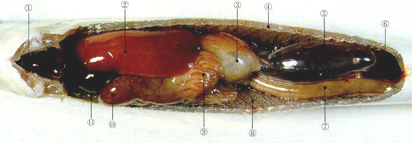
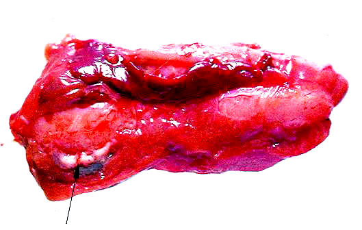
1: heart, 2: liver, 4: peritoneal wall, 7: intestine, 8: peritoneum, 9:pyloric ceca, 10: Stomach, 11: Gall bladder, |
--- The largest accumulation of entodermal tissue in the body
--- Primarily of entodermal tissue
Added by the mesodermal component
--- a. Capsule (Mesothelium) with reticular connective tissue
b. Blood vessels
# Blood of intestine and spleen enters into liver
(1) Location
--- The largest gland located anteriorly within the pleuroperitoneal cavity
--- Contacting with stomach
--- Associated with peritoneum, intestine, spleen, swimbladder and pancreas
(2) Lobulation of liver
a. Unilobe or no lobulation
--- Cyclostomi
Ayu\ Rainbow trout\ Eel\ Pike (Esox)\Mackerel\ Swellfish
b. 2 or 3 lobes
--- Chondrichthyes and many teleosts(2)
Tuna, codfish and flounder(3)
c. Polylobes
--- Polylobes in Mebaru, Urumeiwashi
--- Irregular lobes in Cyprinus carpio
d. Number of hepatic lobes of farmed teleosts
(3) Color of liver
--- Generally, reddish brown
--- Wild fish
--- Variation in color
--- Individual and species differences
--- From yellow to dark brown to gray
--- Diseased state
--- Dark reddish
--- High-fat foods
--- Tinge of yellow
--- Barometer of nutrition and health
(4) Size of liver
--- Associated with species, foods, season or sex
--- Hepatosomatic index
--- Weight of liver/ Body weight X 100
--- Chondrichthyes 10 - 20%
--- 33% in Yumejame (a deepsea species)
Osteichthyes 1 - 2%
--- Exception
--- 4 - 11% in Crucian carp, Samma, Umazurahagi, Kusahugu
--- ? in Cod
Man 2%/ Dog 3%
--- Male < Female (x1.5 - x2.0) in fish
--- Indices in farmed species
--- Below 1% or above 2%
--- How different between sex ?
(5) Blood supply and distribution within the organ
--- Dual supply
--- Several parts of digestive tube
--- Portal vein
<--- Spleen
Dorsal artery
--- Hepatic artery
============> Interlobular vein and artery
--- Sinusoidal capillary within lobule
--- Central vein in lobular center
--- Intercalated vein
--- Hepatic vein
---> Heart
--- Liver in the carp
--- 2 main lobes
--- Longitudinally separated by sagittal septum(Septum sagittale)
--- Sagitall septum
--- A part of dorsal mesentry
--- Passing-through of vessels entering into liver
--- Between 2 lobes
--- The intestine originates
--- Middle furrow or Sulcus medianus hepatis
--- Considered as boundary line separating of 2 main lobes
--- Terminated in the liver center usually
--- Occasionally passing through the liver center
---> 2 separate livers with paired structure
--- Intestinal furrow or sulci intestinales
|
|
|
|
(1) Right lobe (lobus principalis dexter)
--- Gall bladder lobe
--- Surrounding the gall bladder
--- Fovea vesicae biliaris on lobe surface
--- A space in which the gall-bladder is located
--- Gall bladder lobe
--- a. Ventrolateral lobe(Lobus ventrolateralis)
b. Caudal lobe(Lobus caudalis)
--- Rudimentary
(2) Left lobe (lobus principalis sinister)
--- Spleen lobe
--- Surrounding the spleen
--- Impressio lienalis on lobe surface
--- Tied-off from the rest of that organ by intestinal loops
--- Formation of secondary lobe
a. Angular part (Pars angularis)
--- Between the 5th and 8th loops and ventral wall
--- The appearance of a 7
--- Cauda partis angularis
b. Islet part (Pars insularis)
--- Surrounded by the 5th loop
|
|
|
|
|
|
2. Gall bladder 膽囊
--- No gall bladder in dolphins
No gall bladder in Syumokuzame, Nokogiriei and one species of Houbou
(1) Location
--- a. Many of chondrichthyes
--- Embedded within liver
b. Most of osteichthyes
--- Between liver and intestine
--- Opening at the beginning of intestine
(2) Shape and color of the bladder
--- Spherical
--- Flounder\ Swellfish\ Kiankou
Ovoid
--- Mackerel\ Tuna\ Japanese horse mackerel
Slender cord-shaped
--- Mackerel\ Katsuo\ Azi\ Mutsu
--- Yellowish brown or greenish
--- Greenish color suggesting the presence of biliverdin
--- Associated with green liver disease in mullet, parrot fish and red seabream
--- Importance of bile color among species ?
(3) Function
--- a. Emulsification of fat
b. Digestion of proteins
--- Precipitation of certain proteins
3. Pancreas 膵臟
--- The same origin as the liver
--- a. Compact pancreas(Pancreas compactum)
--- Compact only in the elasmobranchii\ dipnoi\ some siluroidei
b. Diffuse pancreas or disseminated pancreas(Pancreas difussum or disseminatum)
--- Diffusely distributed organ
Generally along the portal veins(vv. portae)
--- Appearance at the beginning of portal vein from the intestinal serosa
---> Sometimes penetrated into the liver
--- Hepatopancreas
--- Sufficiently well-developed mesentry between the intestine and liver
---> Concentrated between the epithelial layers of the mesentry
--- Then, appearing a number of thickenings of the intestinal wall
<--- Primary anlagen
<--- Entodermal origin
|
+-- Unite
---> 3 secondary anlagens
--- Fusion
---> Sac-shaped\ externally prominent structures
Located between the stomach and midgut
--- 3 secondary anlagens
--- a. 2 ventral small ones
--- Near the liver anlage
---> Contact with the duct of the liver
--- Left\ right sides of the intestine
---> Exocrine pancreatic tissue
b. A dorsal large one
--- Placed on median plane
--- 3 clusters of primary anlagen
a. A single median cluster
---> Endocrine portion
b. One on each side of the body (2 lateral clusters)
---> Exocrine portion
(1) Location
--- As follicles between esophagus and blind sac-like projection in end of midgut in cyclostomi
--- Separated tubular compact organ with 1 or 2 lobes attached around border between
stomach and intestine
--- Compact organ on dorsal wall of digestive tibue in dipnoi and on dorsal side of spiral intestine
in Siragansu
--- Diffuse organ in many of teleosts which is dificult to discern with naked eyes.
a. Individual organ
--- Anguilla\ Silurodei(Sheatfish)\ Pike(Esox)
--- Easily discernible pancreas
b. Around liver
--- Crucian carp\ Japanese horse mackerel
c. Hepatopancreas
--- Carp\ Flatfish\ Tilapia\ Sea bass\ Red sea bream
d. On the pyloric ceca
--- Salmon\ Trout
--- Within the connective tissue
e. Around digestive tube
--- Mugill
--- May be surrounded by fat tissue within the mesentry
f. Splenopancreas or around spleen
--- Loach\ Angel fish
g. Around Islet of Langerhans
--- Mackerel
(2) Shape of compact pancreas
--- Chondrichthyes
--- Variation with species
--- 2 lobes with small one around intestine and with large one on dorsal
side of stomach
(3) Openings of pancreatic duct
--- A duct system separated from bile duct system even in the liver having
hepatopancreas
--- The begining of intestine
Hepatopancreas
--- Pouring independently from that of common bile duct
(3) Islet of Langerhans (Insula pancreatica)
|
|
|
[Islets of Langerhans in carp, arrow] |
--- Covered by thin exocrine layer
--- Location of endocrine islets in teleostei
--- a. Within the isolated pancreatic tissue/ Around liver
b. Discrete near gall bladder (around Ductus choledochus)
--- As small bodies surrounded by connective tissue
c. Within the spleen
![]()
I BACK I RETURN I NEXT I SUMMARY I Any Questions? I
![]()