![]()
I BACK I RETURN I NEXT I BOTTOM I SUMMARY I
![]()
Endocrine organs 內分泌 組織 또는 臟器
1. Pituitary gland
2. Saccus vasculosus
3. Epiphysis
4. Urophysis spinalis
5. Thyroid gland
6. Ultimobranchial gland
7. Thymus
8. Pseudobranch
9. Adrenal system
10. Corpuscles of Stannius
11. Island-cells of the pancreas
12. Digestive tract as endocrine organ
![]()
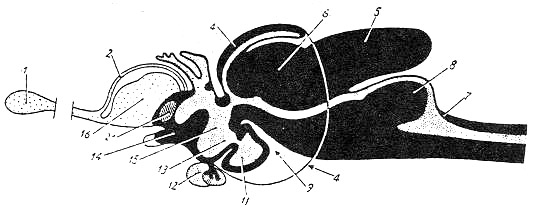
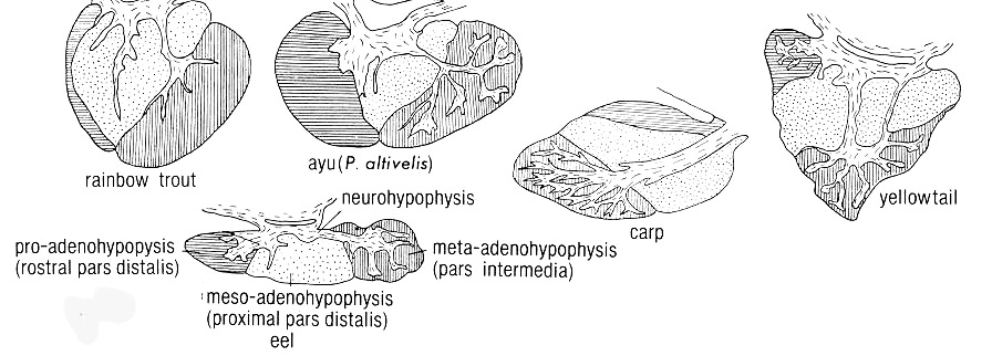
Pituitary (gland) 腦下垂體(12)
|
|
|
15: diencephalon |
Syns. : Hypophysis (cerebri)\ Glandular pituitaria
--- Found in all vertebrates
[Embryological aspects]
--- Ectodermal origin
a. The ventral wall of the 3rd ventricle (ie., diencephalon)
--- Neural tissue
--- Neurohypophysis (Posterior lobe)
b. The roof of the oral cavity
--- Ectodermal epithelium
--- Adenohypophysis
--- No Rathke's pouch in the teleostei and amphibia
1) Location
--- Beneath the diencephalon of the brain
--- Suspended from hypothalamus
--- In a pit on the dorsal surface of the parashenoid bone
--- Called as Sella turcica (Turkish saddle)
--- Attached to the brain by means of its stalk
--- Lobus inferior lobe of infundibulum
--- Directly anterior to the pituitary
--- Covering the pituitary dorsally
Saccus vasculosus
--- Posterior to the pituitary
2) Composition
(1) Adenohypophysis
--- a. Anterior lobe
b. Transitional lobe
c. Intermediate lobe
(2) Neurohypophysis
--- Posterior lobe
|
|
|
|
3) Functions
--- Reference to histology
--- Secretion of Prolactin\ ACTH\ TSH\ GTH
![]()
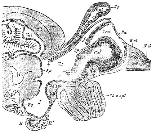
Saccus vasculosus(11) 血管囊
|
|
|
|
1) Shape
--- An unpaired, vesicular organ with high degree of vascularization
Considerable variation in its size and vascularization among the species of teleostei
# paired in elasmobranchii
2) Location
--- Closely posterior to the pituitary
--- An evagination of the wall of the infundibulum
--- A thin walled structure
--- High degree of vascularization
--- Externally apparant
Outer surface covered by the meninges
3) Functions
--- Still subject to much discussion
--- a. 1910, a kind of depth-indicator
--- Not proven
b. 1953, a gland influencing the composition of the cerebrospinal fluid
c. A chemoreceptor and/or a pressoreceptor
1970, regulation of the pressure of the brain liquor by absorption
A correlation between the abundance of the granula in the coronet cell and pregnancy
in the elasmobranchii(Etmopterus niger)
![]()
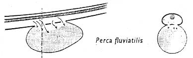
Epiphysis(3) 上生體\ 松果體
# Pineal organ (gland)
|
|
|
|
|
|

--- Projected dorsally from the epithalamus of diencephalon
1) Shape
--- Tube-shaped in teleostei and elasmobranchii
--- A distal thickening
A somewhat-broadening base
===> a. Pineal stalk
b. A pineal vesicle(End vesicle)
--- Slightly infolded epithelium
A pineal lumen
2) Location of end vesicle
--- Projected dorsally from the epithalamus of diencephalon
a. Anterior to the telencephalon
--- Coming into contact with the roof of the cranial cavity
b. Above or posterior to the telencephalon
c. Above the mesencephalon
3) Composition
a. A connective tissue sheath covering the entire organ
b. Internal ependymal lining
c. Lumen communicating with the 3rd ventricle
4) Functions
--- Generally sensitive to light
--- Certain similarity in histology to the retina of the eye
--- Mainly influencing the cycle of daily activity
![]()
Urophysis spinalis p277\Fig 217
|
|
|
|
|
|
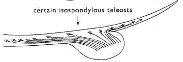
# Synonyms
--- Caudal neurosecretory system 尾部神經分泌系
Neurophysis spinalis caudalis
Caudal hypophysis
Urohypophysis
--- Recognized only in teleosts and elasmobranchs
--- A ventral thickening of the spinal cord
--- At about the beginning of the urostyle or in the region of the last caudal vertebrae
--- More strongly vascularized than the dorsal portion of the spinal cord
--- a. Ventral portion
--- Rather strong vascularization
b. Dorsal portion
--- Neurosecretory cells
--- Three structural types of urophysis
a. Continuous transition f개m the spinal cord to the urophysis
b. An incomplete membrane separates the spinal cord from the neurosecretory tissue
c. A constriction in the stransition zone which partially separates the urophysis from the rest of the
spinal cord
A filum terminale posterior to the urophysis
--- Functions
--- An organ deposits which releases materials produced in the neurosecretory
cells located in the spinal cord
--- Considerable variation of opinions as to the function
a. Osmoregulatory or not ?
--- Capillary network
---> Caudal veins
---> Kidney
---> Increase in retention of Na+
b. Contraction of smooth muscle
c. Possible relationship with the active and abrupt motion of caudal portion
![]()
Thyroid gland 甲狀腺
|
|
|
|
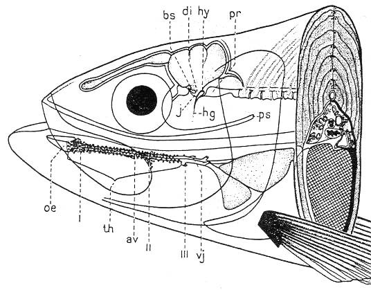
--- The floor of the gill-basket
--- More or less large accumulation of vesicles or follicles
--- Developing out of entoderm from the floor of the gill basket
--- Anlage in the region of the first 2 gill slits
--- Migration into the proximity of the 1 and 2 branchial arteries and the ventral aorta(aorta
ventralis)
Thyroid gland in teleosts
--- Generally diffusely distributed in the region of the above mentioned blood vessels, the ossa
basibranchialia and the m. sternohyoideus
--- Blood supply
--- Ventral branches or elongation of the efferent gill arteries
--- No parathyroid gland in fishes
--- Perhaps this gland has taken over the parathyroid function
![]()
Ultimobranchial bodies
--- Vesicular protrusions from the last gill slit
--- postbranchial or suprabranchial bodies
# Suprabranchial chamber or organ, or labyrinth as an air-breathing organ
--- Originating from the region of the gill slits
--- Both sides or ventral to the esophagus near the sinus venosus in Teleostei
--- Confined to the space between the gill basket, the esophagus and the pericardium
1) Batoidee(Rays\ Sharks)
--- Paired and bilaterally arranged to the pericardium
Selachoidei(Sharks)
--- Only formed on the left side of the body and located on the dorsal wall of the
pericardium, in a nook between the basi- and the hypobranchial of the last branchial
arch
2) Teleostei
--- On both sides(Pike - Esox, and salmonidae) or ventral to the esophagus near the venous
sinus
--- Discrete organ in all calsses of vertebrates except for agnatha(Missing)
--- Fused to the thyroid in mammals
--- Functions
--- Calcitonin
--- Inhibiting the breakdown to the bone tissue by the osteoclasts
--- Prevention of a hypocalcemia
--- Antagonist of the parathormone
Thus, different from that of the parathyroid in mammals
--- Origin of parathyroid
---> The 3rd\ 4th gill-slits of pockets
![]()
Thymus 胸線
|
|
|
|
|
|
--- On the sides of the gill basket in all fishes except adult agnatha
--- A cortical\ a medullary portion
--- Usually difficult to separate from each other
--- Strongly vascularized
Variation in shape and size
--- On the superior edge of the gill cover towards the opercular cavity in Teleostei
![]()
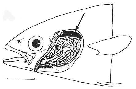
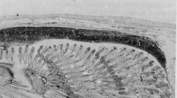
Pseudobranch 위새
--- An endocrine function
--- On the inner side of the gill cover or in about the same region on the base of the skull
--- Often deeply embedded in the surrounding tissue
--- Functions
--- The exchange of CO2
![]()
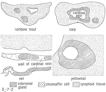
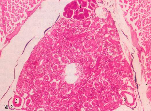
The adrenal system 副腎系
|
|
|
|
|
|
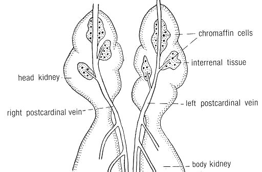
--- Not a unitary adrenal gland
--- 2 different types of tissues
--- More or less regularly distributed
--- The region of the kidneys and the larger blood vessels
===> a. Interrenal system
b. Suprarenal system
<== Very different origins
1. Interrenal organs
--- Developing out of the peritoneum
--- Mesodermal origin
--- Anlage
--- Spliting off on both sides of the mesentery between mesonephros
--- A frequently-interrupted system of nodules
--- Extending along the entire kidney, from the pronephros to the caudal end of the
trunk kidney
--- Considerable variation in shape and size
a. Located in the lymphomyeloid tissue of the head kidney (Trout)
--- Isolated from the cardinal veins and vascularized by blood vessels of its own
b. Some of the interrenal tissue in most other teleostomi
--- Assuming the form of a thicker or thinner glandular layer surrounding the posterior
cardinal vein and its tributaries
--- Functions of interrenal organ
--- Hypertrophy during sexually-active phase
--- Gonadectomy
--- During season of maturation
---> Inhibition of the hypertrophy
Sexually-matured individual
---> A rapid reduction of tissue
--- No affect on the pituitary
--- Hypophysectomy
--- Atrophy of the interrenal organ
A reduction of its enzyme content
2. Suprarenal organ
--- Homologous to the adrenal medulla
--- A completely different origin from the interrenal organ
--- Developing out of the nerve tissue of sympathetic ganglia
--- The nodules of this organ
--- Paraganglia
--- Chromaffine- or phaeochrome system
--- Adrenaline and noradrenaline
--- Chromaffin cells located outside of the organ
--- a. Pronephros (Head kidney)
b. Interrenal tissue
c. Between cardiac muscle cells (Petromyzon)
<=== Chromaffin cells
--- Complete missing of chromaffin cells in Brachidanio rerio(cyprinidae)
--- The organ in agnatha\ elasmobranchii\ chondrostei\ teleostei
--- Located in the region of the head kidney or shortly posterior to it
The organ in the salmonidae\ esocidae
--- The interrenal and suprarenal organs
--- Separated from each other
![]()
The corpuscles of Stannius
|
|
|
|
--- Only found in holostei\ teleostei
--- Usually located somewhat dorsally in the middle line of the caudal portion of the kidney,
where the right and left kidneys come into contact with each other
--- Pale, spherical piles of cells
--- Alveolar structure
--- Developing exclusively out of the mesonephros
--- Formation of nodules
--- Completely separated from mesonephros
--- Differentiation into corpuscles
--- Functions
--- Removal of the corpuscles
--- Alteration in the level of Na+, K+ and Ca2+ in the blood
--- Wound, esp., burns
--- Strong activation of the corpuscles
--- Strong vascularization
--- Endocrine organ
--- No formation of corticosteroids
![]()
The island-cells of the pancreas 膵內分泌細胞
--- Generally not a compact organ
--- Usually diffuse
--- Running along the veins of the hepatic portal system and bile ducts into the liver
--- Covering the wall of the intestine and the surface of the liver
--- Endocrine portion of pancreas in teleostei
--- a. Region of the exocrine pancreas
b. Mesentery and spleen
c. Hepatic portal veins
d. Gall bladder
![]()
The digestive tract as endocrine organ
1) Stomach
--- Gastrin\ Serotonin
--- Stimulation of the secretion of intestinal and pancreatic juices
2) Small intestine
--- Hormones from the small intestine
--- Influencing the secretion of bile and pancreatic juice and the activity of the stomach
--- Little known about these hormones and the histological and cytological bases of their
production
--- Particularly nothing known concerning this type of hormonal activity in fishes
--- Enterochromaffin cells with basally located granula and preenterochromaffin cells found in
latimeria(Intestine\ Also rectal gland)
--- Serotonin
--- Stimulation of the peristalsis of the intestine as one of the functions
Secretion of cholestokinin-pancreozymin found in the intestine of Chimera monostrosa
(holocephali)
The entire gastro-intestinal endocrine system
--- a. Probably primarily important for the organ in which it is locatd
b. Sensations as those of hunger\ satiation
![]()
The urogenital tract as an endocrine organ
--- Testis\ Ovaries
--- Hormones
--- Directly or indirectly related to reproduction
--- Controlled by hormone from the pituitary gland
--- Gonadotrophic hormones(GTH)
--- Stimulation of the secretion of the specific sex hormones
--- The specific sex hormones
--- Only synthesized in the presence of the spermatogonia or the oogonia
--- Thus, probable production of those cells
a. Interstitial tissue or organ
--- The Leydig cells
--- Also described in teleostei
--- Morphologically similar in all vertebrates but not easy to find
--- Found in Lampetra fluvitalis\ Latimeria\ Scyliorhinus\ Chimera\ Belone\ Lebistes\
Gasterosteus\ Coris\ Solea\ Salmo gairdneri\ Blenniidae\ Tilapia\Clupea sprattus
--- The location of Leydig cells in teleostei
--- Not always found in the same position as are in other vertebrates
--- a) The wall of the testicular tubuli ie., lobule-bound in many species
b) The intertubular space(Salmo salar\ S. gairdneri)
--- Formation of clusters
--- True interstitial cells
b. Sertoli cells
--- Formation of a kind of supporting tissue between the spermatogonia and the spermatocytes
--- As nurse cells
--- Involved in the production of steroid hormone in sharks and teleostei
c. The epithelium of the ductus efferentes(Xenentodon cancila, Beloniformes)
--- Also producing the steroid hormones
d. The theca interna of the primary follicles in the female lampreys
--- Probable secretion of hormones
![]()
The kidneys as an endocrine organ
--- Glandular epitheloid or juxtaglomerular cells
--- Walls of the afferent arterioles (middle tunic)
--- Developing out of smooth muscle cells
--- Equivalent to the juxtaglomerular cells of mammals
--- No production of hormones
--- Production of renin, a proteolytic enzyme
---Angiotensin
--- blood pressure
Urine formation
![]()
I BACK I RETURN I NEXT I TOP I
![]()